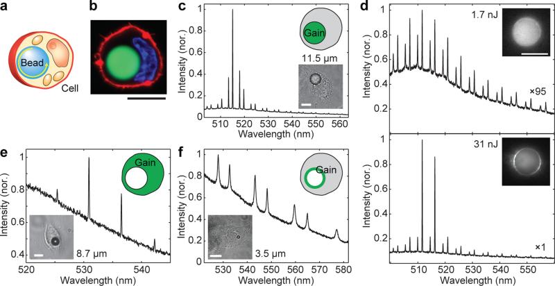
Figure 3. Three different types of solid intracellular microcavities.
a, Illustration of a bead inside a cell. b, Confocal fluorescence image of a HeLa cell containing a polystyrene bead (green), nucleus (blue) and plasma membrane (red). c, Laser emission from a fluorescent polystyrene bead inside a cell. d, Emission spectra and images (insets) of a fluorescent polystyrene bead below and above lasing threshold (3.2 nJ). e, Laser output from a 8.7 μm non-fluorescent BaTiO3 bead embedded in a cell that contains CMFDA dye in its cytoplasm. f, Spontaneous emission from a 3.5 μm BaTiO3 bead coated with Alexa 488 dye below laser threshold. Scale bars, 10 μm.

