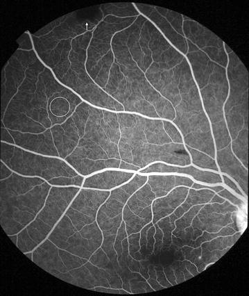
Fig. 13.

Grade 1 peripheral capillary non-perfusion (CNP). Superior retina (right eye). Individual areas of CNP are <1/3 disc area. 1/3 disc area is shown as a white circle. Note that the dark lesion (arrow) is a haemorrhage, and not CNP. Haemorrhage masks background fluorescence and has rounded edges, while CNP has a geographic boundary. Punctate focal leak is visible at the macula
