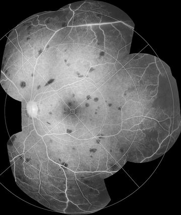
Fig. 17.

Grade 3 peripheral capillary non-perfusion (CNP). Montage of FA images (left eye). Individual areas of CNP are >1 disc area (superior and temporal quadrants), but do not extend into zone 2 nasally or zone 1 in other quadrants. Bays of CNP cut across large vessels and ghost vessels may be visible (e.g. temporal quadrant)
