Fig. 28.

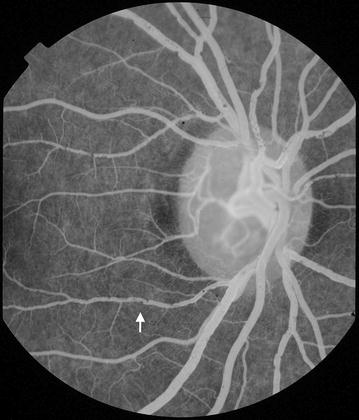
Intravascular filling defects (IVFD). 20° image (right eye). In this image IVFD are prominent in vessels at the disc (white arrow). The venules appear to be affected much more severely than corresponding arterioles

Intravascular filling defects (IVFD). 20° image (right eye). In this image IVFD are prominent in vessels at the disc (white arrow). The venules appear to be affected much more severely than corresponding arterioles