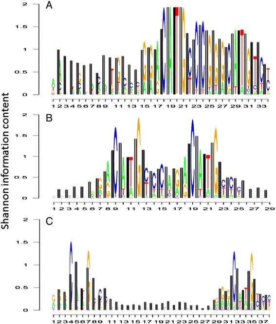
Fig. 2.

TRX-logos plots describing Shannon information content regarding base sequence and intrinsic DNA flexibility on the DNA backbone for the TP53 tumor suppressor binding site. Lighter shaded bars indicate more flexible conformational states. Shown here are the TP53 binding signatures collected using two molecular biological approaches, (a) 17 sites deposited on JASPAR using CASTing or cyclic amplification and selection of targets method of Funk et al. [40] Note: this method is now referred to as SELEX (b) 1231 ChIP-seq sites deposited on JASPAR (c) 150 sites isolated by ChIP-seq study of Cui et al. [41] (note: sequences were center aligned to each TF bound fragment and flanked to the longest spacer retrieved
