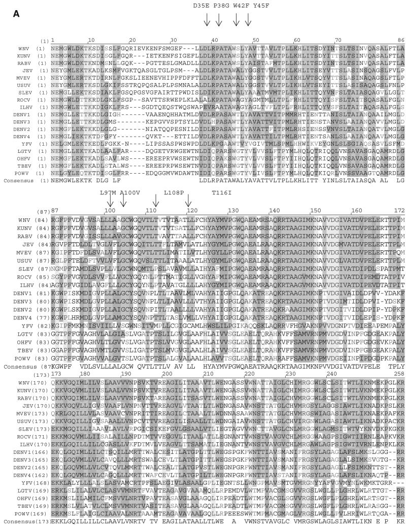
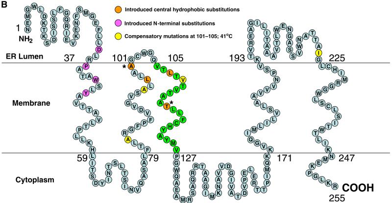
Fig. 1.
Primary amino acid sequence of NS4B proteins from different flaviviruses and predicted secondary structure of WNV NS4B. (A) Complete NS4B amino acid alignments including both tick-borne and mosquito-borne flaviviruses show locations of introduced substitutions in the conserved N-terminal region (D35E, P38G, W42F, Y45F) and central hydrophobic domain (L97M, A100V, L108P, T116I). Virus abbreviations: WNV: West Nile NY99 strain (NC_009942); KUNV: Kunjin (AY274504); RABV: Rabensburg (AY765264); JEV: Japanese encephalitis (NC_001437); MVEV: Murray Valley encephalitis (NC_000943); USUV: Usutu (NC_006551); SLE: St. Louis encephalitis (NC_007580); ILHV: Ilheus (NC_009028); ROCV: Rocio (AY632542); DEN1: dengue 1 (NC_001477); DEN2: dengue 2 (NC_001474); DEN3: dengue 3 (NC_001475); DEN4: dengue 4 (NC_002640); YFV: yellow fever Asibi strain (AY640589); LGTV: Langat (NC_003690); OHFV: Omsk hemorrhagic fever (NC_005062); TBEV: Tick-borne encephalitis (NC_001672); POWV: Powassan (L06436). (B) A topological model was constructed based on the consensus prediction (ConPredII) hydrophobicity plotting program (Arai et al., 2004). Five transmembrane domains were predicted, and amino acids 106–126 represent the primary transmembrane domain (green). Predicted locations of introduced N-terminal substitutions are shown in purple while introduced central hydrophobic substitutions are shown in orange. Positions of additional compensatory substitutions (A83S, A95T, A100V, V110A, I224V) observed in the P38G/T116I/N480H virus incubated at 41 °C are shown in yellow. *A100V and T116I substitutions were introduced as central hydrophobic substitutions but also represent compensatory substitutions following introduction of the P38G substitution.


