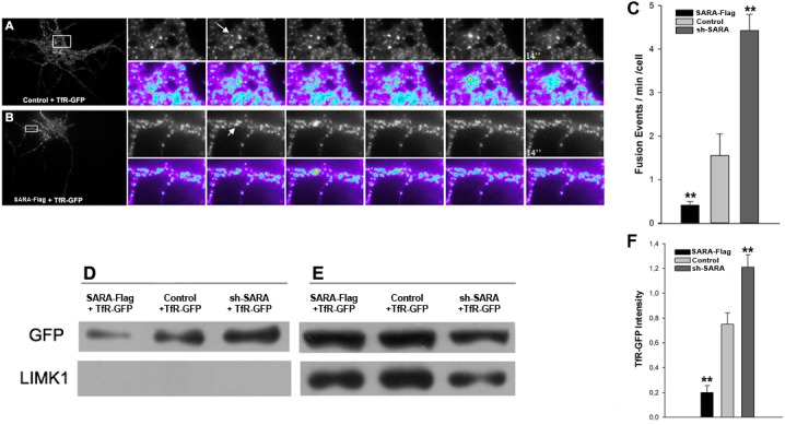
Fig 6. SARA suppression perturbs somatodendritic protein delivery.

Time lapse images by TIRFM showing fusion events of vesicles to the plasma membrane in neurons of 7 DIV, transfected with TfR-GFP plus HcRed (A) or SARA (B). Images were taken every 2 seconds for at least 2 minutes. The graphic shows a significant reduction of fusion events at the somatodendritic plasma membrane in cells transfected with SARA plus TfR-GFP and significantly more fusion events in sh-SARA transfected neurons (C; n = 17 neurons; **p<0.001). TheTfR-GFP cell-surface protein increases after SARA silencing. CHO-K1 were transfected with TfR-GFP alone or plus SARA-Flag or sh-SARA. The cultures were incubated with the membrane-impermeable sulfo-NHS-biotin at 4°C to prevent endocytosis. Then the cells were harvested and biotinylated proteins were collected with avidin-agarose beads (Pierce). Membrane (D) and intracellular (E) proteins were isolated by centrifugation and analyzed by Western Blot with the indicated antibodies. Absence of cytosolic contamination was corroborated with anti-LIMK1 antibody. Graph showing the increase of TfR-GFP fluorescence intensity relative to total protein load (labeled with Tubulin; F) in SARA suppressed neurons respect to control or SARA overexpressed neurons. Data are means of three independent experiments.
