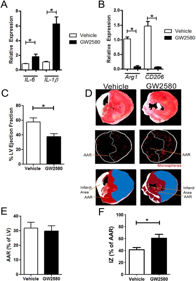
Fig 3. CD206+ M2 macrophage depletion reduces LV ejection fraction but not infarct size 2 weeks post MI in MAFIA mice.
A. mRNA expression relative to GAPDH of M1 markers IL6, IL1B, and (b) M2 markers Arg1 and CD206 respectively within the heart 2 weeks post MI (n = 4–6 animals per group). C. Determination of ejection fraction based on pressure-volume loop measurements vehicle or GW2580-treated MAFIA mice 2 weeks post MI. (* p<0.05. n = 11–13 animals per group). D. Upper panels: representative MAFIA heart sections after TTC staining. Middle panels: representative MAFIA heart section under fluorescent microscope allowing the detection of coloured microspheres distributed in the perfused area. Lower panels: Infarct areas are represented as white, non-perfused area-at risk (AAR) of infarction (dark red) and perfused areas (blue) are highlighted. The area at risk corresponds to the non-perfused area. E. AAR/LV ratio quantification in MAFIA mice 2 weeks post MI using coloured microspheres. F. IZ/LV ratio quantification in MAFIA mice 2weeks post MI using TTC staining and coloured microspheres (* p<0.05. n = 11–13 animals per group).

