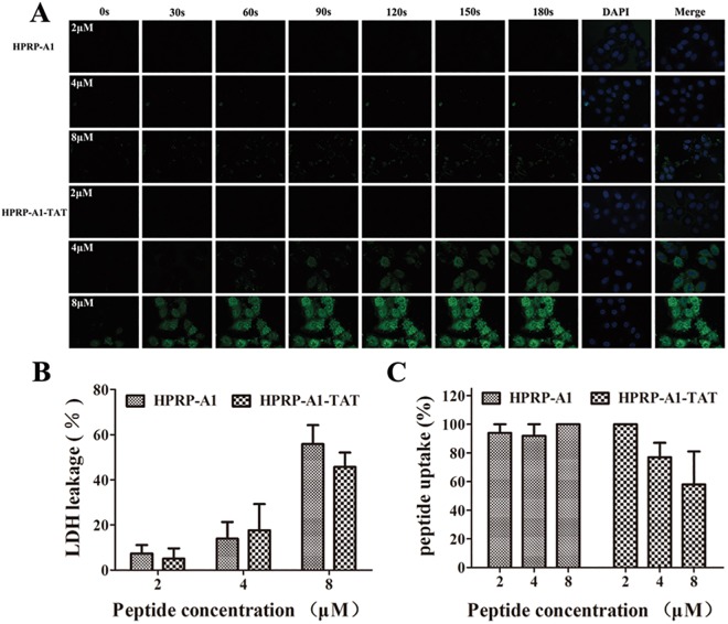
Fig 2. Cellular uptake and interaction between peptides and cell membranes.
(A) The fluorescence time profiles of interaction between peptides and membranes. HeLa cells were incubated with different concentrations of FITC-labeled HPRP-A1 and HPRP-A1-TAT at concentrations of 2, 4, and 8 μM. Images (400× magnification) were captured by laser scanning confocal microscopy every 30 s from 0 to 180 s. Green, FITC peptides; blue, 4,6-diamidino-2-phenylindile-stained nuclei. (B) LDH leakage assay. HeLa cells were incubated with HPRP-A1 and HPRP-A1-TAT at 2, 4, and 8 μM for 1 h and LDH assayed. (C) Cellular uptake of peptides, measured by flow cytometry. After incubation for 1 h at 4°C, HeLa cells were incubated with FITC-HPRP-A1 or FITC-HPRP-A1-TAT peptides for 1 h. The cells were those cultured and treated with peptides at 37°C were used as controls. Data are presented as the mean ± SD of three independent experiments. LDH, lactate dehydrogenase.

