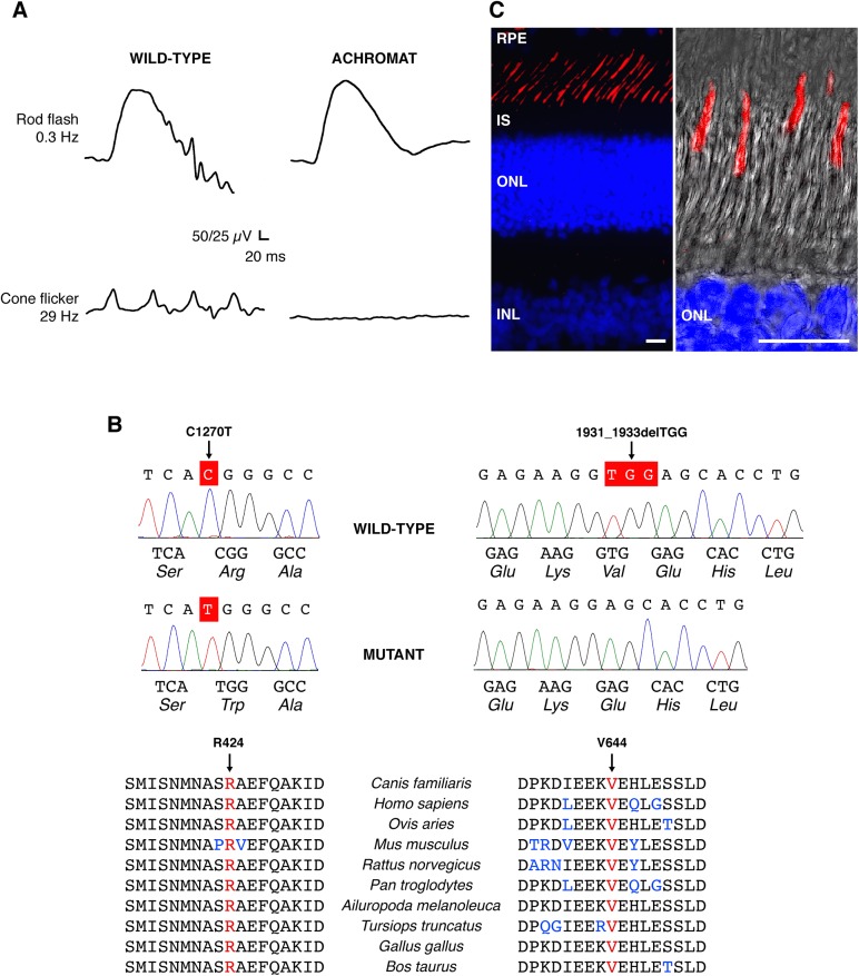
Fig 1. CNGA3-mediated channelopathy in the canine model: clinical manifestation and genetic basis.
(A) Scotopic and photopic ERG responses obtained from a 5-month-old CNGA3-R424W-affected dog showing normal rod flash response, and absence of cone-mediated flicker responses compared to the age-matched wild-type control in ERG responses evoked under dark-adapted (rod flash, 0.3Hz) and light-adapted (cone flicker, 29 Hz) conditions. See also S1 Fig for behavioral vision testing under bright- and dim-light conditions. (B) DNA sequencing chromatograms of the wild-type and CNGA3 spontaneous mutants showing the C1270T transition in exon 7, and the 1931_1933delTGG deletion. Both residues are highly conserved among vertebrate genomes. Canis familiaris—dog; Homo sapiens—human; Ovis aries—sheep; Mus musculus—mouse; Rattus norvegicus—rat; Pan troglodytes—chimpanzee; Ailuropoda melanoleuca—panda; Tursiops truncatus—dolphin; Gallus gallus—chicken; Bos taurus—cow. (C) Immunohistochemical representation of CNGA3 protein in the wild-type canine retina. CNGA3 is exclusively expressed in the outer segment of cone photoreceptor cells (labeled in red), shown also in a high-magnification confocal photomicrograph (right) representing a Z-stack of maximum projection images taken at 0.5 mm intervals created with Leica LAS-AF software (Leica Microsystems, Inc.). Cell nuclei were stained with DAPI. RPE: retinal pigment epithelium; IS: inner segment; ONL: outer nuclear layer; INL: inner nuclear layer; Scale bars: 10μm.

