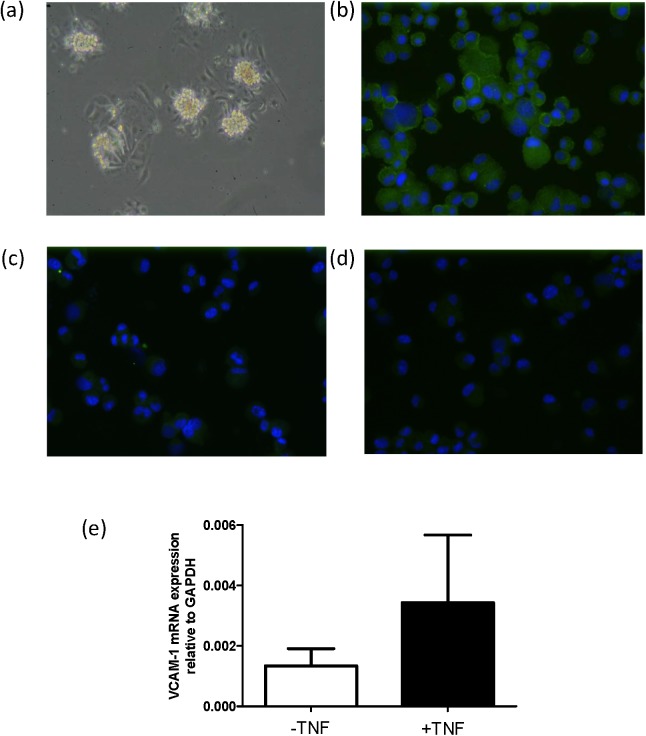
Fig 3. VCAM-1 is expressed in primary mouse podocytes.
(a) Phase-contrast view of glomerular outgrowths of isolated glomeruli from mouse kidney, 2 days after seeding in podocyte-specific medium. Immuno-staining of cytospots of the ICAM-2 negative fraction after ICAM-2 re-beading, detecting nephrin (b), while the endothelial marker Tie2 (c) is not detected. Panel (d) shows background staining with control rabbit-IgG antibodies (and does not detect anything). Antibodies that specifically bound to the cells are stained in green. Nuclei were stained in blue with DAPI. (e) VCAM-1 mRNA expression in mouse primary podocytes in the absence (-) or presence (+) of TNFα, as determined by RT-qPCR. Data are presented as mean values +/- sd, n = 3 from three independent experiments.

