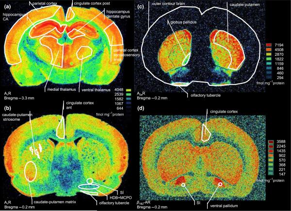
Figure 2.
Representative autoradiograms showing the receptor distribution in the baseline condition and how the regions of interest were defined. (a,b) [3H]-DPCPX (8-cyclopentyl-1,3-dipropylxanthine) binding to adenosine A1 receptors at two coronal sections. (c) [3H]-ZM 241 385 binding to adenosine A2a receptors. (d) [3H]-DHA (dihydroalprenolol) binding to adrenergic β1- and β2-adrenergic receptors. The shape and size of some brain regions varied markedly between the three individual sections analysed at each anatomical level. For example, the globus pallidus often appeared much bigger than it does in the –0.2 AP level shown in (c). CA, cornu amonis; SI, substantia innominata; HDB, horizontal limb of diagonal band; MCPO, magnocellular preoptic nuclei.

