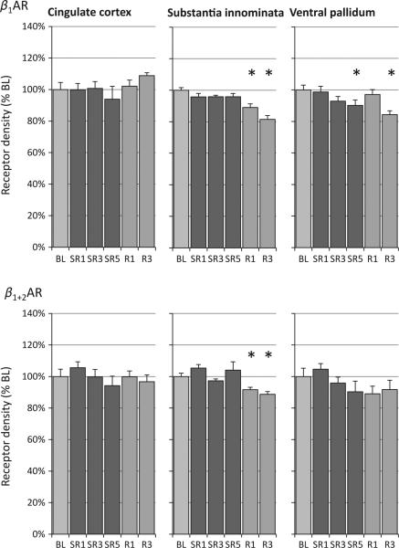
Figure 5.
Relative changes of β-adrenergic receptor (β-AR) density during sleep restriction (SR1, SR3 and SR5) and recovery sleep days (R1 and R3) compared to baseline (BL). Chronic sleep restriction decreased β1-AR density (top panel) in the substantia innominata and ventral pallidum; this reduction remained at R3. No changes were found in the anterior cingulate cortex. A similar pattern was seen with a ligand that binds to both β1-AR and β2-AR (bottom panel; see text for details). See Table 1 for analysis of variance (anova) and linear contrast statistics; the asterisks (*) indicate statistical significance of post-hoc comparisons to BL (P < 0.05, n = 8).

