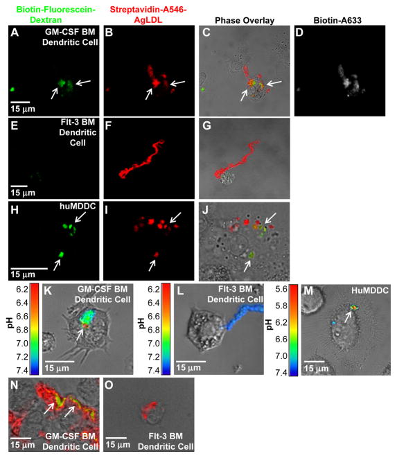
Figure 2. GM-CSF BM DCs use exophagy to degrade agLDL but flt-3 BM DCs do not.
(A–J) GM-CSF BM and flt-3 BM DC lysosomes were loaded with biotin-fluorescein-dextran via overnight incubation. DCs were subsequently incubated with streptavidin and Alexa546 dual-labeled agLDL for 90 min, fixed and permeabilized. Colocalization of dextran (green) with the aggregate (red) indicates areas of exocytosis in mature GM-CSF BM DCs (arrows, A–C). A short pulse of Alexa633-biotin prior to fixation confirmed that the agLDL was still contained in an extracellular compartment (D). No lysosome exocytosis to areas of contact with the aggregate was observed in flt-3 BM DCs (E–G). Lysosome exocytosis to an extracellular agLDL containing compartment was confirmed in mature huMDDCs (arrows, H–J). (K–M) GM-CSF BM and flt-3 BM DCs were incubated with CypHer 5E, a pH sensitive fluorophore, and Alexa488, a pH insensitive fluorophore, dual-labeled agLDL and the pH surrounding the aggregate was measured. When mature GM-CSF BM DCs interacted with the dual-labeled agLDL, regions of low pH could be seen at the contact sites (K). No acidification was observed in aggregates in contact with classical DCs (L). Regions of low pH were also seen in contact sites between mature huMDDCs and agLDL (M). (N and O) GM-CSF BM and flt-3 BM DCs were incubated with Alexa546-agLDL for 1 hr, fixed and stained with filipin to indicate free cholesterol. (N) Cholesterol accumulation can be seen in compartments containing agLDL in an immature GM-CSF BM DC (arrows). (O) No cholesterol generation was observed when flt-3 BM DCs were incubated with agLDL.

