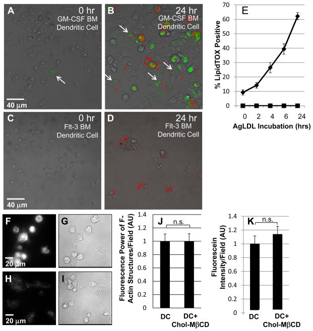
Figure 3. GM-CSF BM DCs incubated with agLDL form foam cells but flt-3 BM DCs do not.
Immature GM-CSF BM DCs and flt-3 BM DCs were incubated with Alexa546-agLDL (red) for 0, 2, 4, 6 and 24 hrs. Cells were then fixed and stained with LipidTOX (green) to assess neutral lipid droplet formation. (A) Few LipidTOX positive non-classical DCs (arrow) are seen in the absence of agLDL. (B) GM-CSF BM DCs incubated with Alexa546-agLDL for 24 hrs form foam cells. Arrows indicate DCs in contact with agLDL containing LipidTOX positive droplets. (C) No LipidTOX positive flt-3 BM DCs are seen in the absence of agLDL. (D) Flt-3 BM DCs incubated with Alexa546-agLDL for 24hrs are negative for LipidTOX staining and do not form foam cells. (E) Quantification of the number of cells containing LipidTOX positive droplets as a function of agLDL incubation time. Diamonds: GM-CSF BM DCs, squares: flt-3 BM DCs. Error bars represent the standard error of the mean (sem). Data are pooled from 3 independent experiments for GM-CSF BM DCs and 2 independent experiments for flt-3 BM DCs. (F and G) Filipin and DIC image of immature GM-CSF BM DCs treated with 5mM cholesterol-MβCD for 15 min. (H and I) Filipin and DIC image of resting immature GM-CSF BM DCs. (J) Cholesterol loaded and resting immature GM-CSF BM DCs were incubated with Alexa546-agLDL for 60 min in the presence of an ACAT inhibitor followed by fixation, labeling of F-actin with Alexa488-phalloidin and quantification of the average local F-actin intensity per cell. (K) Cholesterol loaded and resting immature GM-CSF BM DCs with lysosomes labeled with biotin-fluorescein-dextran were incubated with streptavidin-Alexa546-labeled agLDL for 90 min in the presence of an ACAT inhibitor, fixed and permeabilized. The amount of biotin-fluorescein-dextran exocytosed was quantified. Data are pooled from 2 independent experiments. Student’s t test n.s. not significant.

