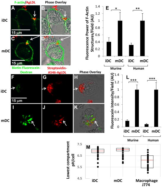
Figure 4. Exophagy is upregulated with DC maturation.
(A–D) GM-CSF BM iDCs and mDCs were incubated with Alexa546-agLDL for 60 min followed by fixation and labeling of F-actin with Alexa488-phalloidin. After 60 min, an enrichment of F-actin was detected near the sites of contact with agLDL in both GM-CSF BM iDCs (A and B) and mDCs (C and D). (E) Quantification of the average local F-actin intensity per cell in both murine and human monocyte derived iDCs and mDCs. (F–H) GM-CSF BM iDCs and mDCs were incubated with biotin-fluorescein-dextran overnight to deliver it to lysosomes. Cells were then exposed to streptavidin-Alexa546-labeled agLDL for 90 min, fixed and permeabilized. Lysosome exocytosis was observed to aggregate containing compartments in both GM-CSF BM iDCs (F–H) and mDCs (I–K). (L) Quantification of the amount of biotin-fluorescein-dextran exocytosed in both murine and human monocyte derived iDC and mDC. (M) GM-CSF BM iDCs, mDCs and J774 macrophage-like cells were incubated with CypHer 5E, a pH sensitive fluorophore, and Alexa488, a pH insensitive fluorophore, dual-labeled agLDL and the pH surrounding the aggregate measured. Quantification of the lowest pH achieved in the compartment at a single timepoint in iDCs, mDCs and J774 macrophage-like cells. The central mark on each box is the median, while the edges of the box represent the 25th and 75th percentiles. Error bars (E and L) represent the sem. * p ≤ 0.05, ** p ≤ 0.01, *** p ≤ 0.001 student’s t test. Data are pooled from 3 independent experiments.

