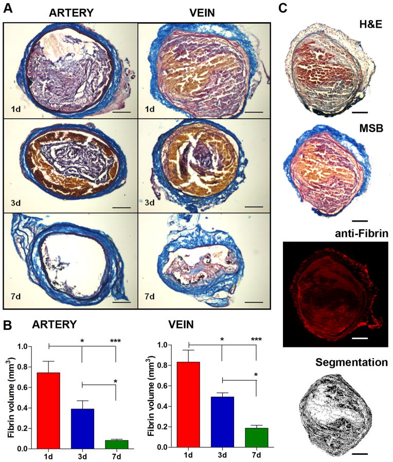
Figure 5. Histopathology.
Representative coronal sections of arterial and venous thrombi stained with MSB showing time-dependent changes in thrombus size and composition (A). Volumetric quantification of thrombus composition revealed a gradual reduction of fibrin content (B). Adjacent histological sections stained with Hematoxylin&Eosin, Martius Scarlet Blue and anti-fibrin antibody showed congruent localization of fibrin, confirming the selectivity of MSB staining for fibrin (C). Color segmentation analysis performed on the MSB-stained section shows fibrin detection. Scale bars: 0.2 mm. Error bars are SEM. ***P<0.001 and *P<0.05, 1-way ANOVA followed by Tukey post-hoc test. n=5/group.

