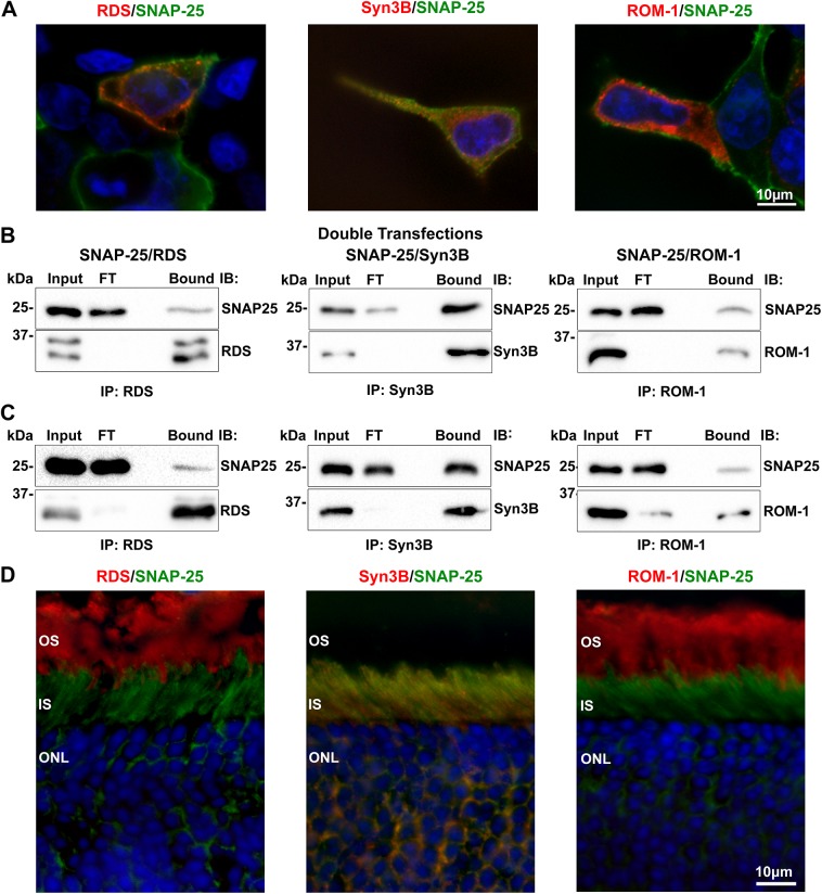
Fig 6. RDS and ROM-1 interact directly with SNAP-25 in vitro and in vivo.
A. HEK293 cells were co-transfected with SNAP-25 (green) and either RDS (RDS-CT, red), ROM-1 (ROM1-CT, red) or Syn3B (Syn3B-529, red) antibodies. B. CHAPS-solubilized cell extracts underwent co-IP for RDS (RDS-CT), Syn3B (mAB 12E5), and ROM-1 (mAB 2H5) antibodies. Resulting blots were probed for SNAP-25, RDS (mAB 2B7), Syn3B (mAB 12E5), or ROM-1 (mAB 2H5) antibodies. C. CHAPS-solubilized WT retinal extract underwent IP and reducing SDS-PAGE/WB with the same antibodies as in B. D. WT retinal sections underwent IF to study distribution of SNAP-25 (green) in the outer retina in relation to labeling with RDS (RDS-CT, red), Syn3B (Syn3B-529, red), or ROM-1 (ROM1-CT, red) antibodies. Scale bars: 10 μm. OS: outer segment, IS: inner segment, ONL: outer nuclear layer. FT: flow through.

