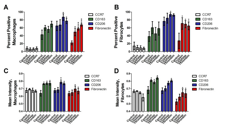
Fig 5. Trypsin, tryptase, and thrombin bias M2 macrophage differentiation towards an M2a phenotype.
PBMC were cultured with MCSF for 7 days to generate M2 macrophages, after which the media was removed and proteases were added to the PBMC for 2 days. Macrophages (A, C) and fibrocytes (B, D) were counted by morphology from representative fields of view. (A) and (B) were performed by eye, while (C) and (D) show analysis of staining intensity. Cells were stained for the indicated markers. Values are mean ± SEM, n = 6. * indicates p < .05, ** p < .01, and *** p < .001 compared to the no-protease control (unpaired two-tailed t-tests).

