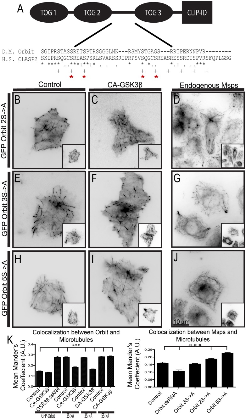
Fig 2. Orbit is phosphorylated by GSK3β in the linker region between TOG2 and TOG3.
(A) Domain structure of Orbit with a Clustal alignment of the GSK3β phosphorylation region. Serines phosphorylated in Homo sapiens (H.S.) CLASP2 denoted by plus marks (+), serines conserved in Drosophila Melanogaster (D.M.) Orbit denoted by red stars. (B-C) GFP-Orbit 2S->A was expressed in cells with a dual expression vector containing tRFP-α-tubulin alone (B) or with CA-GSK3β (C). (D) Endogenous Msps and α-tubulin were stained in cells transfected with 2S->A. (E-F) GFP-Orbit 3S->A is expressed in cells with a dual expression vector containing α-tubulin-tRFP alone (E) or with CA-GSK3β (F). (G) Endogenous Msps and α-tubulin were stained in cells transfected with 2S->A. (H-I) GFP-Orbit 5S->A was expressed in cells with a dual expression vector containing tRFP-α-tubulin alone (H) or with CA-GSK3β (I). (J) Endogenous Msps and α-tubulin were stained in cells transfected with 5S->A. Tubulin images are shown as insets. (K) Changes in co-localization of Orbit (left) and endogenous Msps (right) were measured using the Mander’s coefficient, n = 90 cells from two (endogenous Msps) or three (GFP-Orbit) experiments. *** p<0.0001.

