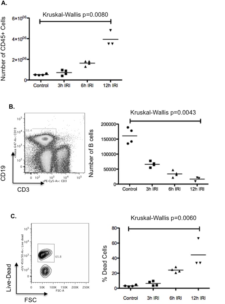
Fig 3. B cells following hepatic IRI.
Following 40 minutes left lobe ischemia, WT mice were allowed to reperfuse for 3, 6 or 12 hours. Mice were then sacrificed and their ischemic left lobes digested. The isolated immune cells were analysed by flow cytometry. There was a significant influx of immune cells (defined as CD45+ cells) into the injured left lobe [Kruskall- Wallis p = 0.0080] (A). B cells were defined as CD3-CD19+ gated lymphocytes. The number of live B cells was found to decrease significantly with time following reperfusion [Kruskall- Wallis p = 0.0043] (B); this was predominantly due to cell death (as defined by positive staining for a live-dead marker) [Kruskall- Wallis p = 0.0060] (C).

