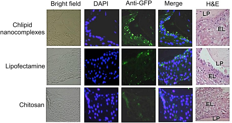
Fig 3. Transfection of cells in a 3D-VEC model.
Cells in the 3D-VEC model were transfected with either chitosan or Lipofectamine 2000 or CNs complexed with pEGFP. At 48 h post-transfection, the tissues were fixed in 10% formalin, paraffin-embedded, and immunostained using anti-GFP antibody and nuclear-stained with DAPI. H&E stained histological (formalin-fixed) cross-sections of VLC-FT are also shown. LP, lamina propria; EL, epithelial layers.

