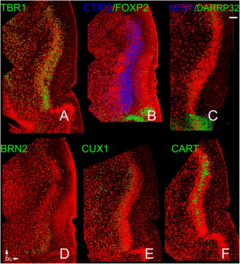
Fig 4. Patterns of neocortical layer markers in the APC.
A) TBR-1 heavily labeled cells in Layer 2 as well as scattered cells in Layer 3. Of the 4 deep layer markers (B,C), only CTIP2 exhibited dense staining. The other three (FOXP2, NOR1 and DAARP32) labeled sparse number in Layers 1–3. The dense staining for FOXP2 and DAARP32 seen at the bottom of the figures sharply demarcates the APC from the more ventral OT. The other three makers exhibited very different patterns: BRN2 staining was found more in the ventral APC (D), CUX 1 in the deeper portions of both Layer 2 and 3 (E), and CART in the middle of Layer 2(F). Scale bar = 200μm. Dorsal to top, lateral to right.

