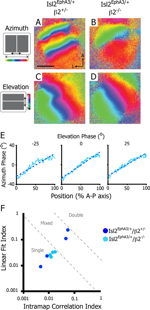
Figure 6.
Disruption of retinal waves eliminates heterogeneity in the azimuth representation in the SC of Isl2EphA3/+ mice. A–B) Intrinsic signal optical imaging of the azimuth representation in Isl2EphA3/+/β2+/− (n = 4) (A) and Isl2EphA3/+/β2−/− (n = 4) (B). C–D) Intrinsic signal optical imaging of the elevation representation in Isl2EphA3/+/β2+/− (C) and Isl2EphA3/+/β2−/− (D). E) Phase plots of the azimuth representation along the anterior-posterior axis of the SC at three isoelevation points from an Isl2EphA3/+/b2−/− SC. F) Map organizations of Isl2EphA3/+/b2+/− (dark blue dots) and Isl2EphA3/+/b2−/− (light blue dots) SCs plotted by two quantitative indices. bar, 0.5 mm; A, anterior; L, lateral

