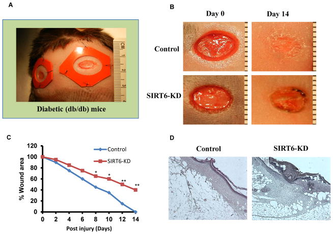
Fig 1. Delayed cutaneous wound healing after SIRT6 knockdown in db/db mice.
(A) Full-thickness skin wounds were created in diabetic db/db mice. (B) Representative photographs showing the macroscopic wound closure on day 14 postinjury from Control and SIRT6-KD diabetic wounds. (C) Microphotographs of wounds was analyzed at indicated time intervals to determine closure in control and SIRT6-KD. (D) Histopathology (H–E staining) of control and SIRT6-KD wound on day 14 postinjury. SIRT6-KD wounds showed less re-epithelialization and granulation formation. A stratified neoepidermis was visible on the edge of the wounds in control, whereas the epithelial tissue was disorganized in SIRT-KD wounds. Representative results from three independent experiments with three-four animals in each group are shown. All values represent the mean ± SEM. *p < 0.05 indicates significantly and **p < 0.01 indicates very significantly higher values observed in SIRT6-KD compared with control wound.

