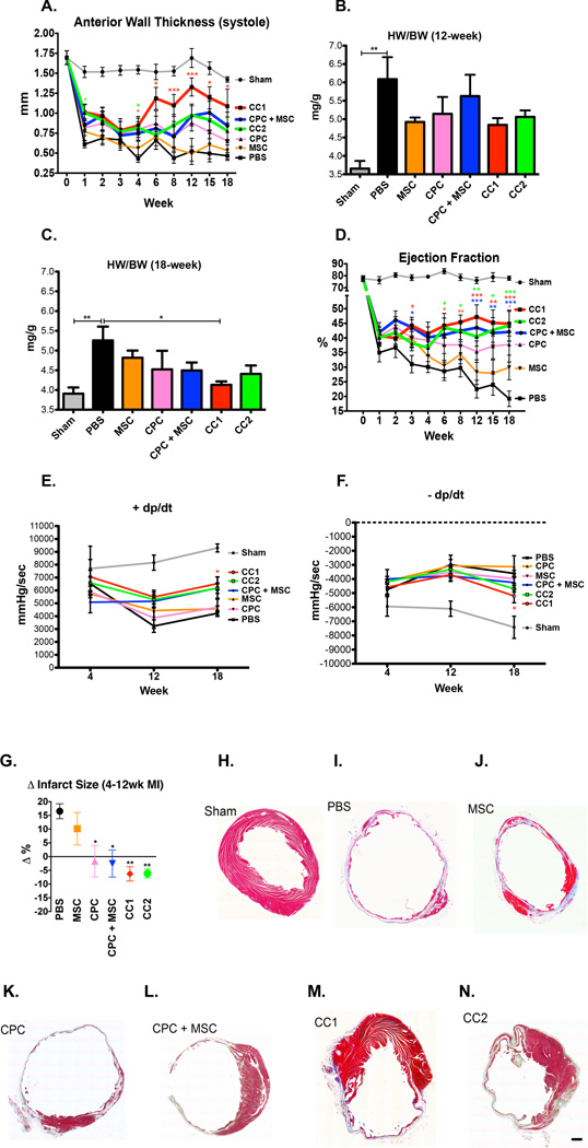
Figure 3. CardioChimeras improve left ventricular wall structure and cardiac function after myocardial injury.
(A) Longitudinal assessment of anterior wall thickness during systole (mm) over 18 weeks. (B) Heart weight to body weight ratio (mg/g) at 12 WPI (C) 18 WPI. Sample sizes of 3–5 mice per group. (D) Longitudinal assessment of ejection fraction (%). (E) Positive and (F) Negative developed pressure over time represented as mmHg/sec at 4, 12 and 18 WPI. (G) Change in infarct size between 4 and 12 weeks time points. P values were determined by one-way ANOVA compared to PBS treated controls. (H–N) Masson’s Trichrome staining and representative images of infarct size and fibrosis in (H) Sham, (I) PBS, (J) MSC, (K) CPC, (L) CPC + MSC, (M) CC1 and (N) CC2. Sample sizes are specified in the Online Table II. All statistical values were determined by two-way ANOVA compared to PBS treated hearts. * p<0.05, ** p<0.01, *** p<0.001. Colors of asterisk(s) correspond to heart group. Scale bar is 250µm.

