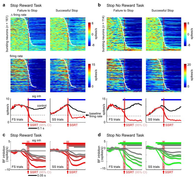
Figure 5. BF neuronal inhibition was similarly engaged regardless of whether stopping was successful.
(a-b) Responses of BF bursting neurons to the stop signal aligned at SSRT, plotted separately for failure-to-stop (left) and successful stop trials (right), and separately for the Stop Reward (a) and Stop No Reward tasks (b). Top row, rate changes in stop trials compared to latency-matched controls. BF bursting neurons were sorted by inhibition latency (white line). Middle row, firing rates in stop trials. Bottom row, population PSTHs aligned to SSRT show that BF neurons were inhibited below baseline firing rates (black dashed line). BF neuronal inhibition was present and time-locked to SSRT in both failure-to-stop and successful stop trials in both SST variants. (c-d) Peri-SSRT inhibition of the five quintiles of BF bursting neurons (mean ± s.e.m.), as defined in Fig.4c-d, in failure-to-stop and successful stop trials. Note that the top three quintiles of BF bursting neurons in successful stop trials are still inhibited 10ms before SSRT. The red shaded areas indicate the 95% CI of SSRT estimate (n=17, 20 sessions).

