
Figure 3.
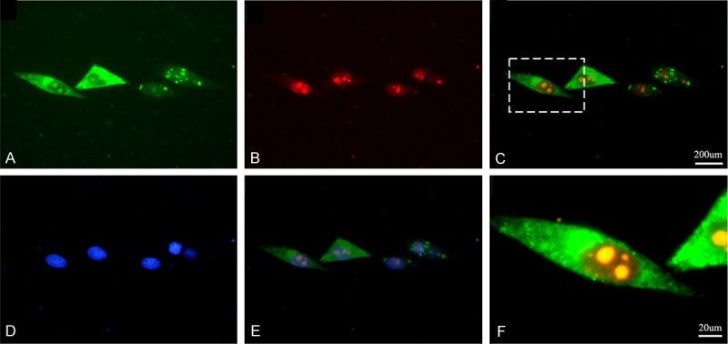
MK and PGRN co-localize in the HepG2 cells in vivo. The MK-RFP and the PGRN-GFP fusion protein expression plasmids were co-transfected into the HepG2 cells, the expression of PGRN-GFP (A) and MK-RFP (B) was detected by fluorescence microscopy respectively. The PGRN-GFP and MK-RFP were overlapped in the HepG2 cells (C), the yellow show the overlap areas, the (F) shows the high magnification of overlapped areas. The cells also stained with Hochest33258 to show the nuclear (D, E).