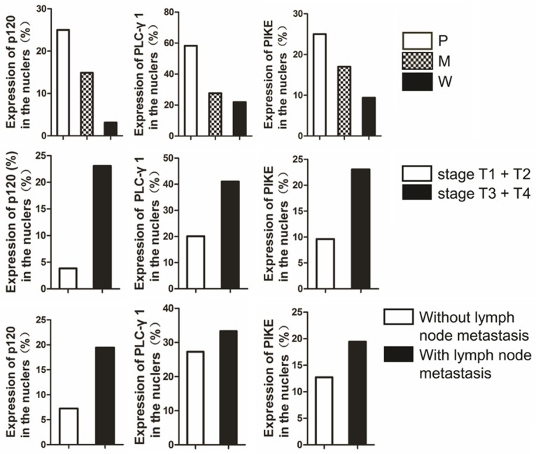
Figure 4.

Nuclear staining of p120, PLC-γ1, and PIKE in OSCC. The percentages of positive cells for nuclear staining of p120, PLC-γ1, and PIKE in OSCC with different levels of differentiation, clinical stages and with or without lymph node metastasis are shown as bar graphs. P, poorly differentiated; M, moderately differentiated; W, well differentiated; N, non-cancerous tissue.
