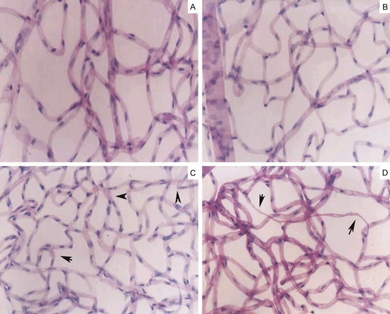
Figure 1.

Retinal histopathology: There are intact vascular network and the uniform diameter of retinal capillary in the normal control group and DM1 group (A, B). In the DM3 group, the vascular net was in uniform distribution and the irregular with tortuous and dilatations of vascular cavity (C, arrow denoted). Acellular capillaries were mainly present in the DM6 group (D, arrow denoted) Magnification × 20.
