
Figure 1.
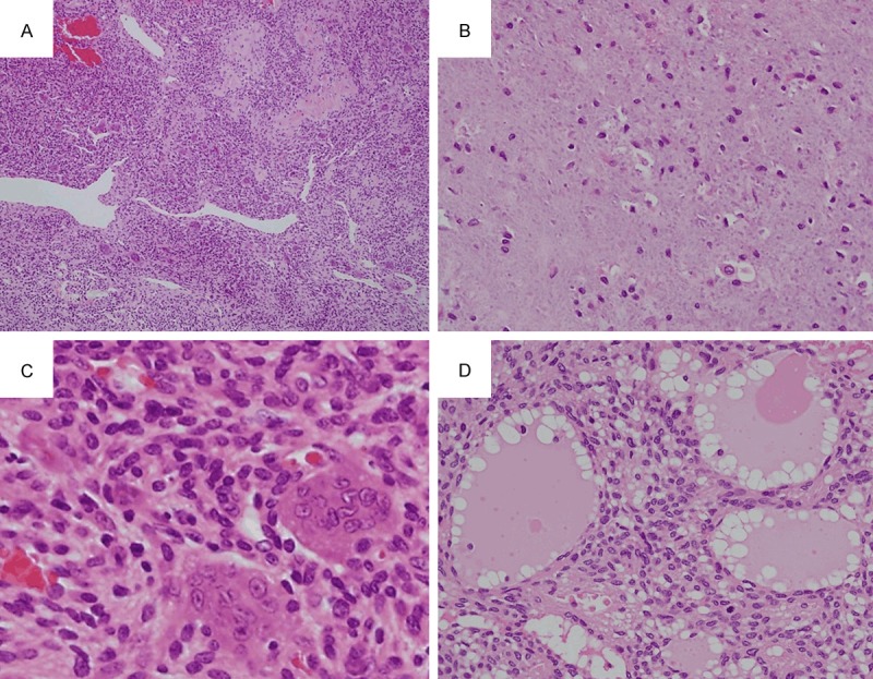
Histological findings. A. Case 8. Monotonous proliferation of tumor cells with a background of prominent microvessels are observed along with the presence of staghorn vessels (×40). B. Case 6. The unique grungy or flocculent calcification is observed (×400). C. Case 8. The tumor cells are spindle-shaped with scant eosinophilic cytoplasm, and bland nuclei with indistinct nucleoli. Osteoclast-like giant cells are also present (×600). D. Case 7. Microcystic structures are observed, in which lymph-like fluid is present (×400).