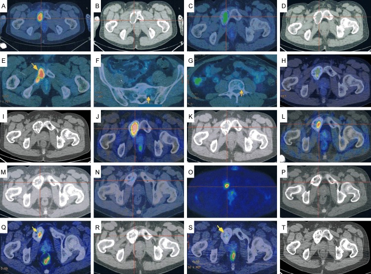
Figure 3.
PET/CT images. A, B: Bone expansion and thinning of the cortical bone in the region extending from the right ischium to the pubic bone. High FDG uptake (SUV max = 9.2) is evident in the soft tissues extending into the bone marrow. C, D: Reduction in size of the tumorous lesion in the right pubic bone (SUV max = 5.5). E-G: Increased FDG accumulation in the right pubic-to-ischial lesions (arrow) (SUV max = 6.6) and new uptake in the sacrum and the first lumbar vertebra (arrows). H, I: The increased FDG accumulation in the right pubic-to-ischial lesions remains unchanged (SUV max = 6.6), whereas the uptake in the sacrum and first lumbar vertebra has decreased. J, K: Worsening of FDG accumulation in the right pubic-to-ischial lesions (SUV max = 9.4) and new uptake in the sternum and fourth lumbar vertebra (SUV max = 1.7). L, M: Diminished FDG accumulation in the right pubic bone lesion (SUV max = 5.0) and sternal lesion (SUV max = 1.4), and disappearance of uptake in the fourth lumbar vertebral lesion. N-P: FDG uptake unchanged in the right pubic bone lesion (SUV max = 4.9), but there is new uptake in the right hip bone (SUV max = 1.8). Q, R: No change in the FDG accumulation in the right pubic bone (arrow) (SUV max = 4.9), but accumulation in the right hip bone lesion has disappeared. S, T: Marked reduction in FDG accumulation in the right pubic bone (arrow) (SUV max = 2.6).

