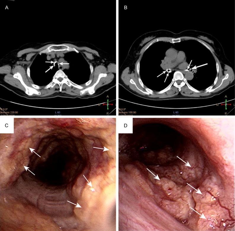
Figure 1.

Images and appearance of TO (A, B) Chest CT scan showing a ring (arrow) of multiple calcified nodules surrounding the tracheal lumen. (C, D) FOB showing dozens of sub-mucosal nodules (arrow) protruding into the lumen of lower half of the trachea and right main bronchi.
