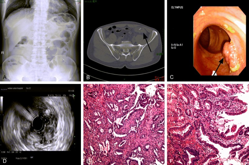
Figure 1.

Preoperative examination results of this triple synchronous colorectal carcinoma patient. A. Abdominal plain film presented with severe flatulence and sporadic air fluid levels; B. Abdominal CT scan with a infiltrative mass and circumferential thickening in descending colon (→); C. Colonoscopy showing a polypoid lesion within sigmoid colon; D. Iso-echoic feature of the sigmoid polypoid lesion by EUS examination; E. Biopsy examination revealing adenocarcinoma of the lesion in descending colon; F. Biopsy examination revealing well-differentiated adenocarcinoma of the polypoid lesion in sigmoid colon; Tissue slides: HE×100.
