
Fig. 5.
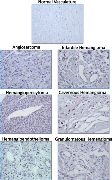
Representastive images of Klf4 staining in normal and vascular tumor tissues. Immunopositivity for Klf4 protein is represented by brown staining. Positive control (left panel) = human intestine; negative control (right panel) = human intestine with no added primary antibody. 400× total magnification for each image