
Figure 2.
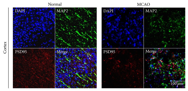
Immunochemical image for confirmation reduced PSD-95 expression in MCAO mouse brain. Immunochemical images showed that PSD-95- (as the post synaptic density protein) positive cells (red) were decreased as expressed in MCAO mouse cortex. Postsynaptic proteins were hardly observed in MCAO mouse brain cortex, whereas the normal cortex was observed evidently. We used 5 rats in each groups for study. Each measurement included 3 repeats per animal. Scale bar = 100 μm, PSD-95: red, MAP2: green, 4′,6-diamidino-2-phenylindole (DAPI): blue, normal: normal control group, and MCAO: reperfusion 24 hr after MCAO injury.