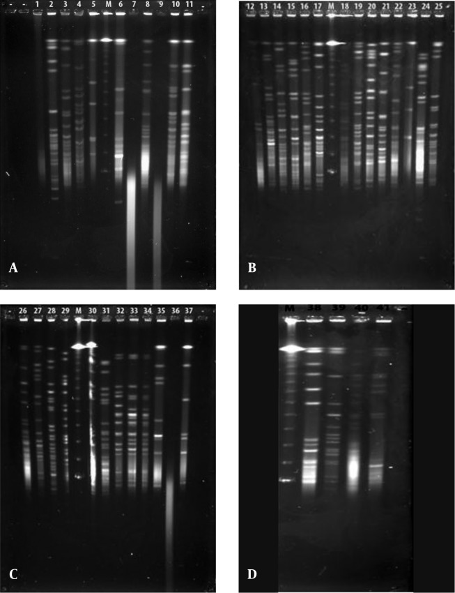
Figure 1. Gel Images of the Forty-One Bioluminescent Strains Obtained From the Pulsed-Field Gel Electrophoresis Analysis With the NotI-HF™ Restriction Enzyme.
A, V. gigantis (1:E-16, 2:E-10, 3:E-11, 4:H-3, 7:S3W46, 8:S3W28, 9:H-16, 10:E-15, 11:H-2, 25:E-14), A. logei (5:Se2Lu45-2), and A. fischeri (6:S2W23) strains; B, V. gigantis (12:SW15, 13:SWLiman , 14:SeLu25 , 15:S2W42 , 16:S2W9 , 17:S3W2, 18:Se2Lu48 , 19:Se3Lu25, 20:FU-10, 21:FU-9), V. lentus (22:FU-7), A. logei (23:U-6), and S. woodyi (24:X-7) strains; C, V. harveyi (26:Se4Lu24, 27:H-15, 28:H-5, 29:H-11), V. orientalis (30:Se4Lu49-2, 31:H-9), V. crassostreae (32:H-7, 33:H-12, 34:H-19), and A. fischeri (35:SW22, 36:E-4, 37:H-18); and D, V. azureus (38:Se4Lu15, 39:H-1, 40:H-14) and P. kishitanii (41:X-8), M: Marker (Lambda Leader, Bio-Rad, USA, 48,5 kb - 1,000 kb).

