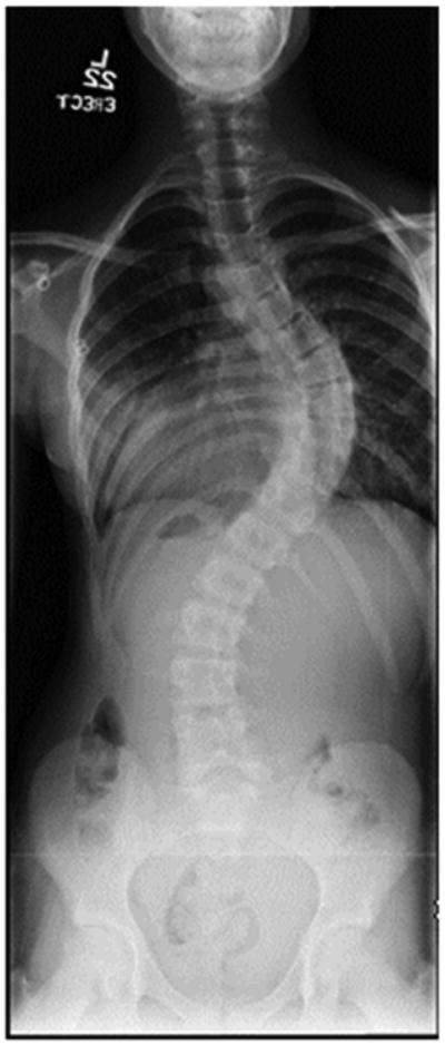
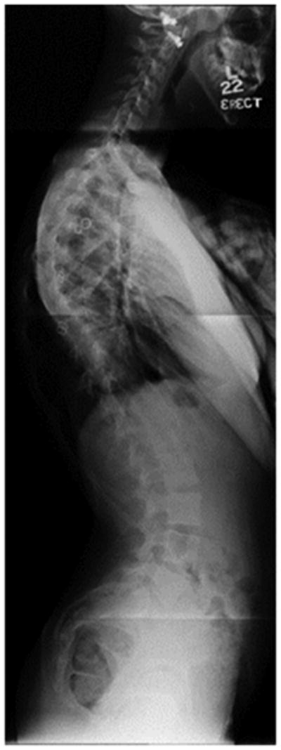
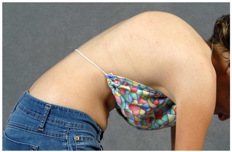
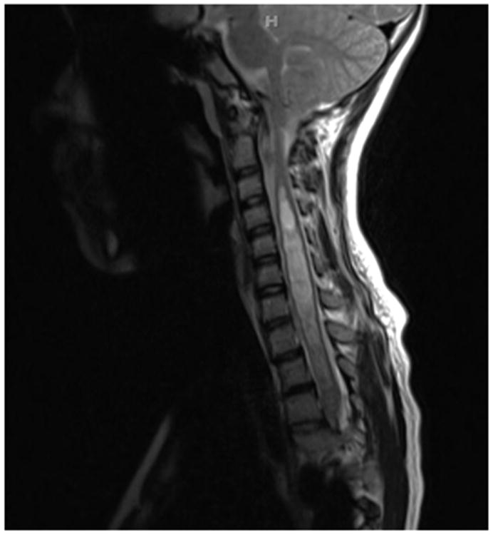
Figure 2.


a - Standing postero-anterior radiograph of a 13 year old girl with “typical” coronal plane findings of adolescent idiopathic scoliosis.
b – Standing lateral radiograph showing proximal thoracic kyphosis, “atypical” for adolescent idiopathic scoliosis.
c – Lateral clinical image of 13 year old girl with proximal thoracic kyphosis.
d – T2-Weighted magnetic resonance imaging of this young woman, showing Chiari-I with syringomyelia.
