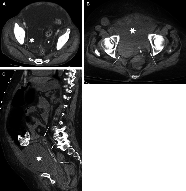
Fig. 8.
Pelvic intraperitoneal fluid vs extraperitoneal fluid. Pelvic fluid is shown in 2 different patients. The patient in A has intraperitoneal fluid. The patient in B and C has extraperitoneal fluid. A axial CT image shows intraperitoneal fluid in the right and left paravesical recesses (asterisks) lateral to the sigmoid colon (S) and superior to the bladder (not shown). B Axial CT image of a different patient shows extraperitoneal fluid in the prevesical space (asterisk) anterior to the decompressed bladder (arrowhead) containing a catheter. The bladder is displaced posteriorly. Fluid extends within the prevesical space (arrows) lateral to the bladder giving a “molar tooth” appearance. C sagittal CT image in the same patient as in B shows extraperitoneal fluid in the prevesical space (asterisk) anterior to the posteriorly displaced bladder (arrows) containing a catheter.

