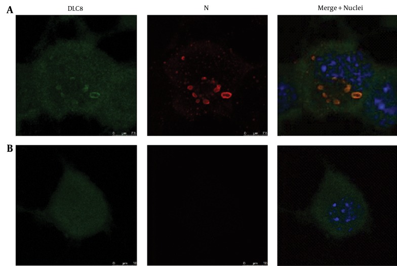
Figure 2. Subcellular Distribution of Dynein Light Chain 8 (DLC8) in the Rabies Virus (RABV)-Infected and Mock-Infected Neuro-2a Cells.
Blue, green, and red fluorescence reflects viable nuclei, host protein DLC8, and N protein of RABV, respectively. A, RABV-infected cells; B, Mock-infected cells. Scale bars = 7.5 μm or 10 μm.

