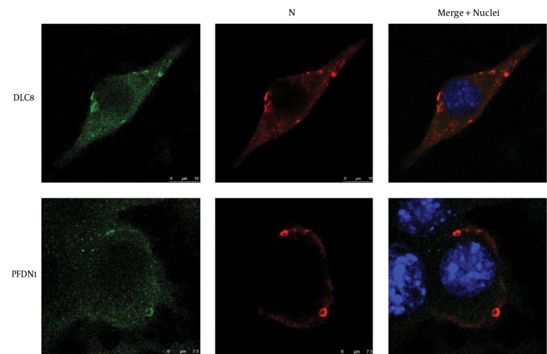
Figure 4. Co-Localization of the Rabies Virus (RABV) Nucleoprotein with Dynein Light Chain 8 (DLC8) and Prefoldin Subunit 1 (PFDN1) in Plasmids Nucleoprotein- and Phosphoprotein-Co-Transfected Neuro-2a Cells.
Nuclei (blue) were stained with 4', 6-diamidino-2-phenylindole (DAPI). PFDN1 and DLC8 are in green and the N protein of RABV in red. Scale bars = 7.5 μm or 10 μm.

