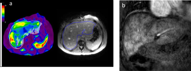
Figure 4.

(a) 31 year old female with a history of pulmonary atresia with tricuspid stenosis, hypoplastic right ventricle, and normally related great vessels, status post neonatal Waterston shunt followed by classic Fontan procedure at age 5 years. A follow-up cardiac MRI was performed to evaluate her Fontan pathway, cardiac chamber sizes, and ventricular function and liver MRE was performed as part of this exam. Mean liver stiffness was 4.0 kPa, well above the accepted normal mean for an adult of 2.3 kPa.
(b) Coronal post-contrast MRI image obtained in the portal venous phase shows diffuse heterogeneous “cloud-like” enhancement of the liver parenchyma, a finding that is seen with liver fibrosis [21].
