
Figure 3.
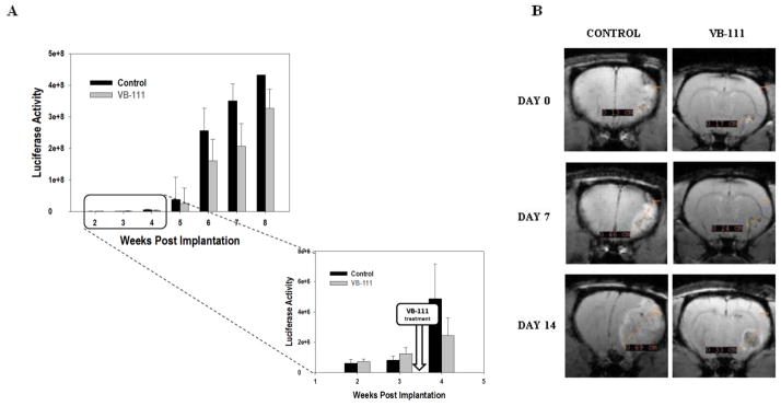
(A) Luciferase emission quantification as a surrogate for tumor volume from nude rats bearing U87MG xenografts (10 animals per group). At week 4 luciferase values separated among the two groups. (B) Representative examples of MRI of treated and control U-87MG bearing rats before (day 0) and after VB-111 treatment (day 7, day 14). The control shows homogenous contrast enhancement, is more solid appearing, and is associated with marked mass effect and midline shift. By contrast, the treated tumor displays central necrosis, minimal mass effect, and a rim of enhancement.