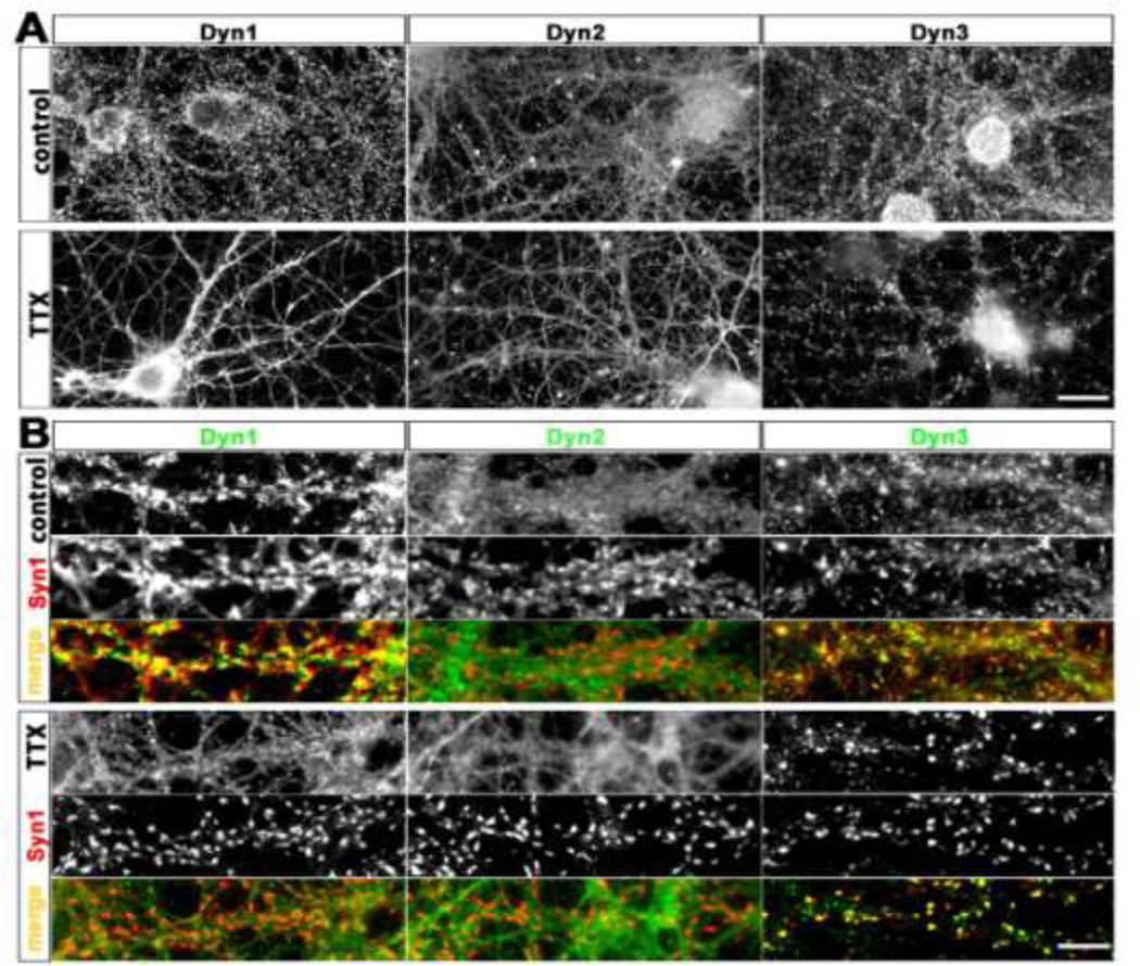
Fig. 1. Distribution of endogenous dynamins in cultured hippocampal neurons before and after chronic suppression of neural activity.
Hippocampal cultures were fixed at 21 DIV and stained using isoform-specific antibodies to Dyn1, 2, and 3, as indicated.
A. Under control conditions endogenous Dyn1 is detectable in a punctate manner along the dendritic arbor, axons and nerve terminals of a 21 DIV hippocampal neuron. Endogenous Dyn2 is detectable in a mostly diffuse pattern throughout all neuronal compartments, and not enriched at synaptic sites. Dyn3 immunoreactivity is characterized by fine puncta present throughout the neurons, with occasional enrichment at some presumptive synapses. Chronic incubation with TTX (1 µM, 14 days) causes an apparent declustering of Dyn1, no detectable change in Dyn2 localization, and a marked accumulation of Dyn3 into larger clusters. Scale bar: 15 µM. B. Selected dendritic regions from hippocampal neurons cultured with or without TTX, immunostained with the presynaptic marker Syn1, together with each indicated dynamin isoforms. Note that the apparent degree of colocalization between Syn1 and Dyn1 increases with TTX, but Syn colocalization with Dyn3 decreases. Scale bar: 8 µM.

