Figure 1.
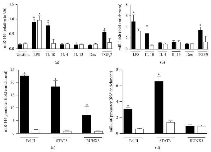
MiR-146b is induced in human monocytes by TGFβ signaling pathway. Human purified monocytes were stimulated for 24 h with 100 ng/mL LPS, 20 ng/mL IL-10, 20 ng/mL IL-4, 20 ng/mL IL-13, 20 ng/mL Dex, or 50 ng/mL TGFβ and (a) miR-146b (black columns) and miR-146a (white columns) levels from total RNA were measured by qPCR in triplicate samples. (b) Cell extracts were subjected to RIP assay using anti-Ago2 or IgG control Abs and levels of miR-146b (black columns) and miR-146a (white columns) were measured by qPCR in triplicate samples. Results are expressed as fold change over control (mean ± SEM, n = 3). (c-d) ChIP assays were carried out on human purified monocytes stimulated or not for 4 h with 50 ng/mL TGFβ (c) or 20 ng/mL IL-10 (d) using anti-Pol II, anti-STAT3, or anti-RUNX3 antibodies. Q-PCR was carried out using specific primers for miR-146b (black columns) or miR-146a (white columns) promoters. Results are expressed as fold change over control (mean ± SEM, n = 3).

