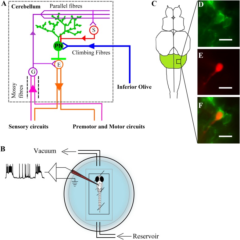
Figure 1. Zebrafish cerebellar circuitry and experimental preparation.
(A) Schematic of the cerebellar circuitry in zebrafish. PN: Purkinje neuron; E: Eurydendroid cell; G: Granule cell; S: Stellate cell. (B) Schematic of the set up for in vivo whole cell recordings in unanesthetized zebrafish larvae. (C) Schematic of the zebrafish larval brain with the cerebellum in green. (D) Mosaic expression of aldoca:gap43-Venus in Purkinje neurons. (E) Patched cell shown filled with sulphorhodamine. (F) Co-localization of sulphorhodamine filled cell with membrane-targeted Venus expression. Scale bar = 10 μm.

