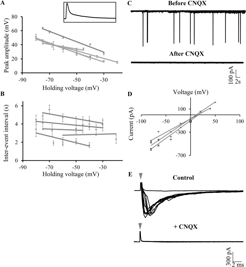
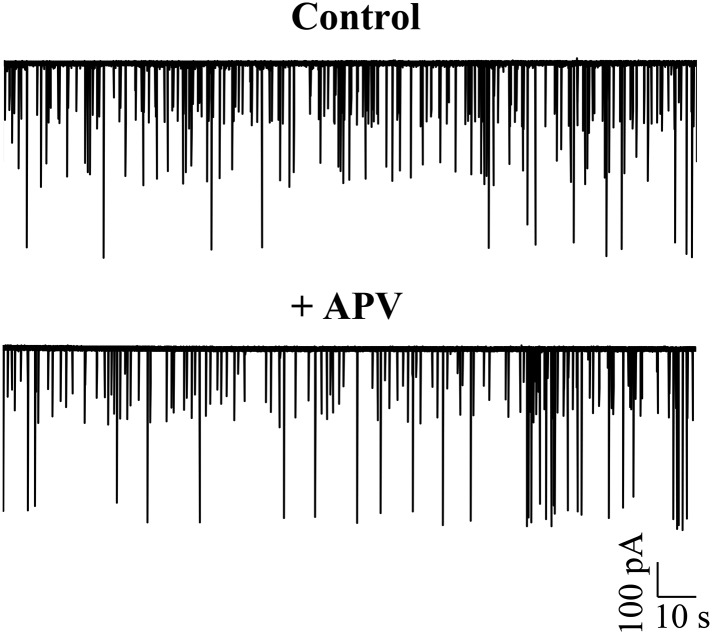
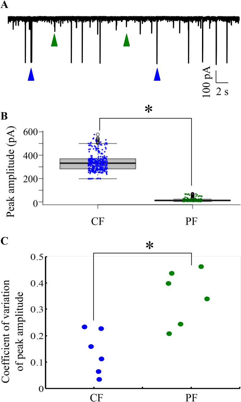
Figure 5. Large amplitude events are climbing fiber (CF) EPSPs mediated by AMPARs.
(A) Mean peak amplitude of large amplitude events as a function of the holding potential. Inset: Expanded trace of a single large amplitude event to illustrate the slow kinetics and large amplitude. Inset x-axis: 400 ms; y-axis: 47 mV. (B) Mean inter-event interval as a function of holding potential. Error bars indicate standard error of mean in A and B (N = 5 cells). (C) Representative trace of a Purkinje neuron recorded in voltage clamp mode at −65 mV before and after application of CNQX. (D) Current-voltage relation of CF EPSCs (N = 3 cells). (E) Representative trace showing all-or-none EPSCs upon stimulation of CFs in the presence of APV and Gabazine (top trace; N = 7 cells). Stimulation at 500 μA resulted in either transmission failure (flat line) or EPSCs of similar amplitudes. In the same cell, all-or-none EPSCs were abolished by the addition of CNQX (bottom trace; N = 5 cells). Grey arrowhead shows stimulus artifact.


