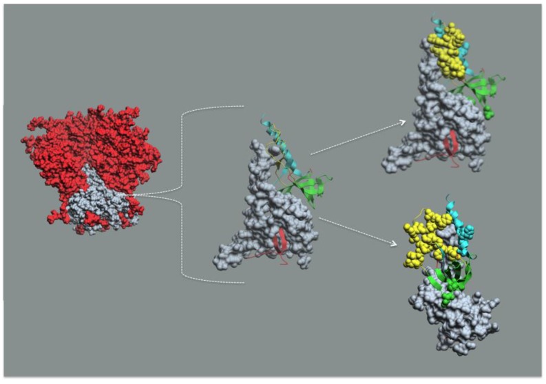
Figure 4.
Epitope Cluster A maps into the gp41 docking site for gp120 in Env trimer mimetic structures. The leftmost structure is the soluble SOSIP Env timer mimetic from PDB:4TVP [95] where gp120 is red and gp41 is gray. The middle figure is a gp41 monomer from PDB:4TVP in gray. The gp41 interactive face comprised of elements from mobile layer 1 (yellow), mobile layer 2 (cyan), the 7-stranded β-sandwich (green), and the N- and C-Terminal extensions (red) of monomeric gp120 shown as ribbon diagrams. The upper rightmost figure is the same as the middle figure except with the N5-i5 and C11 contact residues rendered as cpk structures. The lower rightmost figure is the same as the upper rightmost figure rotated approximately 90°. Note that the N5-i5 contact residues are in mobile layers 1 and 2 (yellow and cyan), whereas the C11 contact residues are in the 7-stranded β-sandwich (green). The viral membrane would be at the bottom of each structure.

