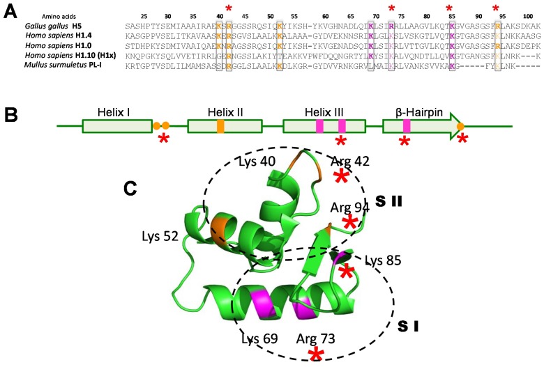
Figure 6.
(A) Amino acid sequence alignment of the WHD regions of several linker histones. The amino acids corresponding to the first and second sites of interaction of this domain with DNA [154] in the chromatosome are highlighted in orange and magenta (respectively). The amino acid numbers refer to their position in the histone H5 sequence; (B) Schematic representation of the secondary structure of the WHD. The sites corresponding to the first and the second histone—DNA interacting domains are in the same colours as in (A); (C) The tertiary structure of the WHD of chicken erythrocyte histone H5 as determined by crystallographic analysis [155], showing the regions and amino acids corresponding to the first (SI) and second (SII) sites of interaction with DNA. The red asterisks highlight the minimal ionic interaction sites that appear to be indispensable for this domain to perform its function in vertebrate linker histones.

