Fig. 3.

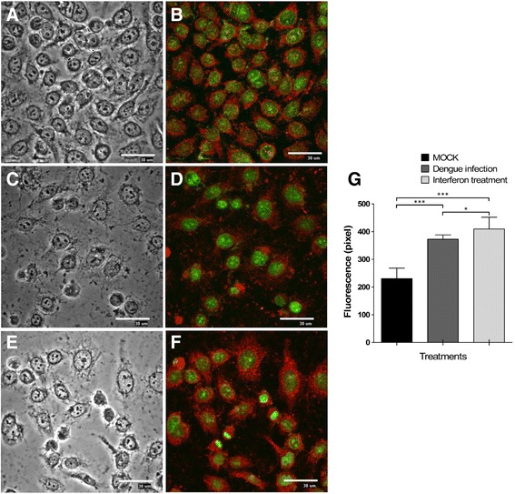
Membrane receptor expression IL28R in C33-A cells. The presence of IL28R1 was determined in C33-A cells: (a, b) Mock-infected. (c, d) DENV-2 infected (MOI = 0.1). e, f Treated with IFN-λ1 (35 ng/ml). Immunofluorescence was detected with anti-IL28R1 and CFL647-conjugated secondary antibody and observed by fluorescent microscopy (40X magnification). g Fluorescence intensities were determined by analysis of three different fields by randomly counting 50 cells using the image analysis software EZ-C1 v.2.3. *p < 0.05, ***p < 0.001. Nuclei were stained with the green fluorescent dye Sybr-14. Bars = 30 μm
